Heart defects
While the foetus is in the womb, abnormalities in the heart may arise, resulting in congenital heart defects.
Heart defects may be relatively simple defects, such as a small hole in the wall between the ventricles of the heart (ventricular septal defect, VSD) or a narrowing of one of the heart's valves. Over time, these defects often disappear as the baby grows. More complex heart defects affect the functioning of the heart and need surgery usually within the first few months of life. Paediatric cardiac surgery is centralized in two hospitals in Sweden, Lund and Gothenburg.
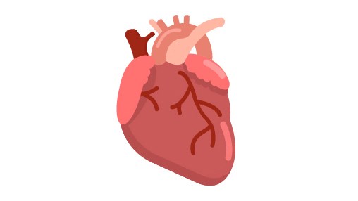
The heart has four chambers, the right and left atria and the right and left ventricles. Blood comes from the body to the right side of the heart and is pumped through the right atrium and right ventricle to the lungs. In the lungs, the blood is oxygenated and then returns to the left atrium of the heart. From there, the blood enters the left ventricle, which pumps oxygenated blood to the body via the aorta. In order to ensure that the blood flows in the right direction through the heart, there are valves between the different chambers of the heart and the major blood vessels.
A heart defect can be detected in various ways. It may have been detected through ultrasound before birth; or the baby may have difficulty oxygenating soon after birth; or the doctor may hear a heart murmur when examining the baby; or the heart defect may not give rise to any symptoms at all. The diagnosis is made by means of ultrasound, a so-called echocardiogram.
Heart defects affect approximately 8 in 1,000 newborns, but only about 2 of these 8 are complex defects. Today, most heart defects can be treated successfully and almost all babies survive, but will need to be followed up during childhood.